Our Hospital
Waiting Area
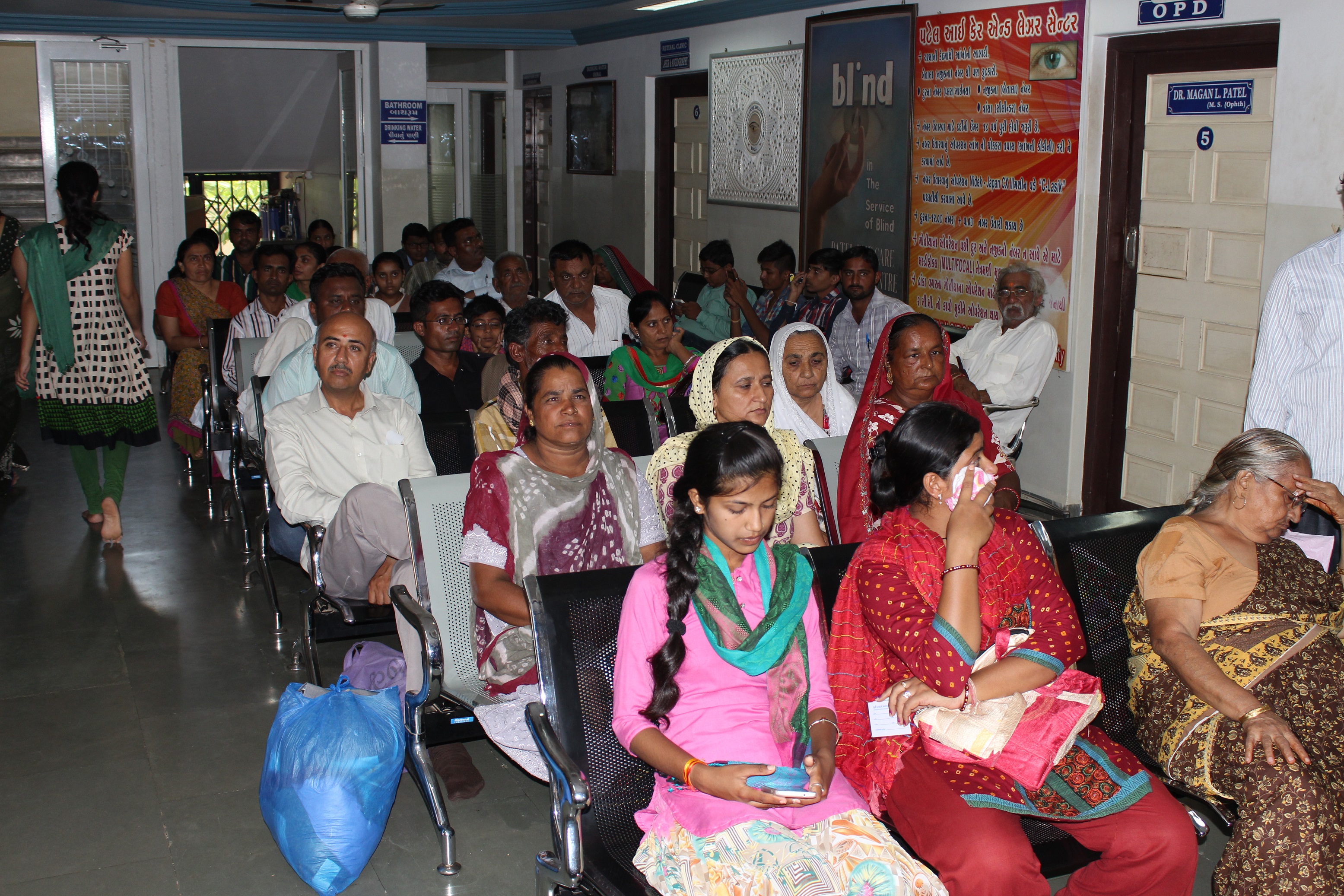 Waiting Room And Among The Most Highly Trafficed Area In a Hospital. So Why aren't hospital waiting Room More Accommodating so We Provide Spacious and well illuminated, well Furnishing Wider Seats And Armrests To Provide Separation Form Stranger, Air Condition Waiting Room First In Kutch.
Operation Theatre
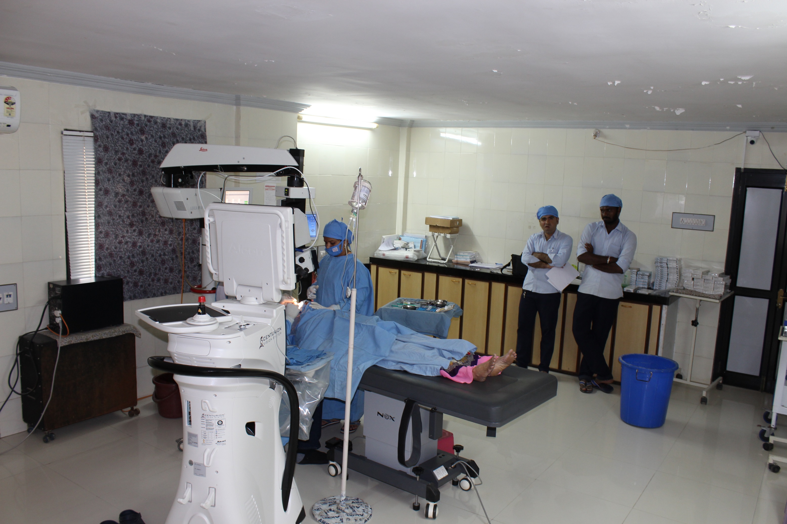
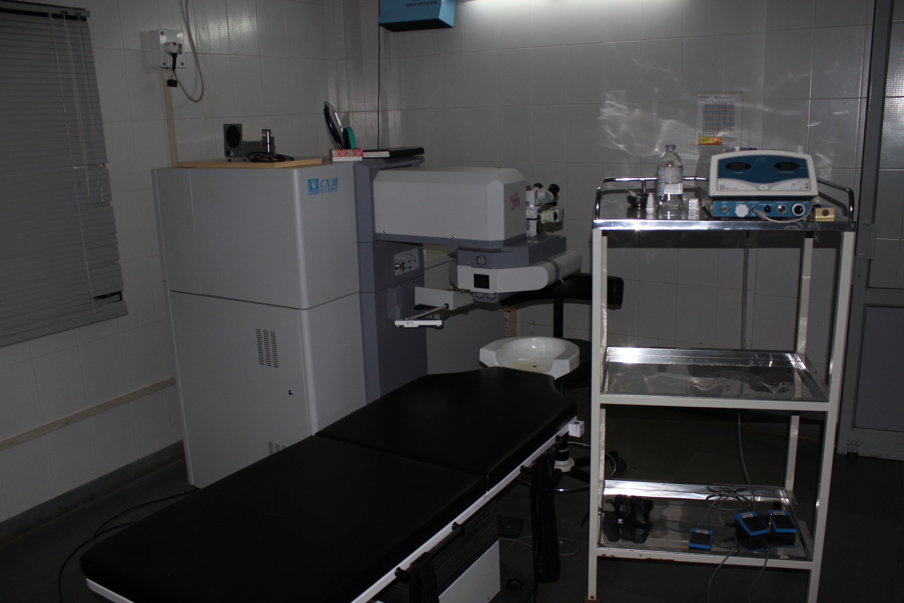 OT is That Specialized Facility of The Hospital When life Saving Or Life Improveing Procedures are carried out On The Human Body by Invasine Mathod Under Strict Aseptic Conditions Is Controlled Enviroment By Specially Trained Personnel To Promote Healing And Cure With Maximum Safety, Comtant And Economy So We Provide World-Class Opration theatre.
Our Hospital Instuments
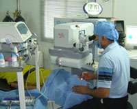
(1) laser machine(Nidek, Japan)
Laser machine with rotational eye tracker and online torsional error detection
*Ideal Candidate For The Lasik Surgery
-patient must be over 18 years of age
-refraction must be stable for a year
-patient should not be pregnant or lactating
-Range of correction should be
-Myopia -1.0 to -10.0D
-Hyperopia +1.0 to +6.0D
-Astigmatism 1.0 to 6.0D
*The Technique-Corneal wavefront guided ( c- lasik)
Aneasthesia-Topical (only putting drops) anesthesia is the choice.
Procedure-the suction ring is fixed. by pressing the foot pedal the microkeratome makes forward pass to cut a corneal flap. The stromal bed is ablated with excimer laser to achieve the desired correction. The flap is reposed .the patient is allowed to blink normally.

(2) microkeratome (Moria,France)
Microkeratome for to create the corneal flap

(3) CENTURION Vision System(Alcon, USA)
World Best Phacoemullsification Machine
CENTURION Vision Systemis designed to optimize every moment of the cataract surgical procedure to improve patient outcomes Provides control and improved efficiency during the minimally invasive cataract phacoemulsification procedure Combines multiple technologies to set new standards in the performance of cataract surgery
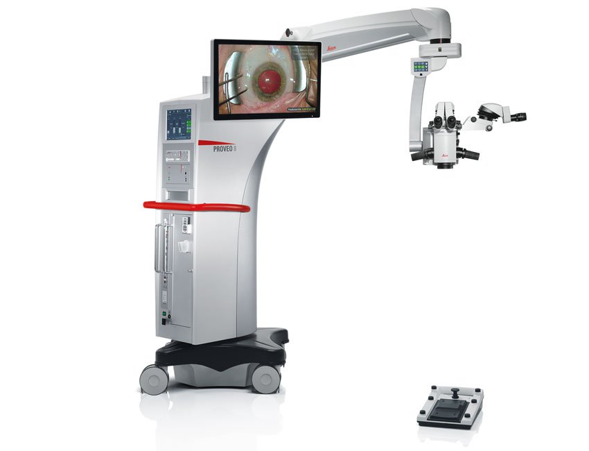
(4) Microscope Proveo 8(Leica,Switzerland)
In the most critical moments of ophthalmic surgery, you need to be able to rely on consistent, uncompromised images, because you can’t treat what you can’t see.
The Proveo 8 ophthalmic microscope goes beyond conventional visualization. Its exclusive optical technology provides you with both constant red reflex and a rich texture view, throughout entire anterior and posterior procedures.
Proveo 8 pushes the boundaries of visualization:
CoAx4 Illumination technology for enhanced view during all cataract surgery stages, including phacoemulsification
FusionOptics technology for better texture view in retina and cataract surgery
(5) VERION Image Guided System(Alcon, USA)

 The VERION Image Guided System: Precise Surgical Planning and Procedures Joining other advanced technologies from Alcon, the VERION Image Guided System is designed to offer improved precision, consistency and control in cataract refractive surgery. The VERION Image Guided System Can help: Minimize data transcription errors Improve clinical efficiency Increase toric and multifocal IOL confidence Ensure surgical consistency Optimize visual outcomes
(6) IOLMaster 500 from ZEISS(ZEISS, Germany)
The ZEISS IOLMaster 500 is the gold standard in optical biometry with more than 100 million successful IOL power calculations to date. With the ZEISS IOLMaster 500 you get a piece of cutting-edge technology. In Patel Eye Care & Laser Centre All Cases Of Cataract IOL Measurement Done By IOL Master 500.

(7) Ellex Yag Laser(Ellex, Australia)
-Capsulotomy PBI
-Laser PBI for Glaucoma Treatment.

(8) Pachymeter(Sonomed,USA)
-To measure the corneal thickness
-Glancoma diagnosis

(9) Diode Green Laser (Iridix,USA)
For The Treatment Of Retinal Disorders
Diabetic Retinopathy
Hypertensive Retinopathy
Macular edema
Central Serous Retinopathy
Retinal hole

(10) Slitlamp Examination(Topcon, Japan)

(11) Fundus Camera
Fundus Photography Fluoresein Angiography
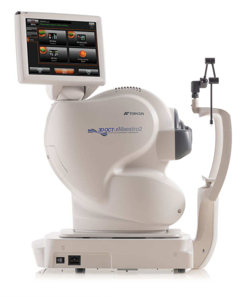
(12) OCT (Optical Coherence Tomography)
Diagnosis For Retinal Disorders
-CME(Cystoid Macular Edema)
-CSME(Clinical Significant Macular Edema)
-Photopic Maculopathy
-CSR(Central Serous Retinopathy)
-ARMD(Age Related Macular Degeneration)

(13) Specular microscope (Topocon japan)
-To count the endothelial cell
-To Detect the endothelial abnormalities.

(14) Auto Ref / Kerato / Tono / Pachymeter TONOREF III
Combination of auto refaractometer, auto keratometer, non contact tonometer and non contact pachymeter High measurement accuracy
Clinically important functions †accommodation measurement and opacity assessment
User-friendly design
Space saving design that is a comfortable and efficient upgrade to your practice

(15) OPD ScanIII(Nidek,Japan)
The OPD-Scan lll is the Five-in-One true refractive workstation combining Wavefront Aberrometer
Topographer
Auto Refractometer
Auto Keratometer
Pupillometer and Pupillographer
Overview summary for opti

(16) Pachymeter TONOPACHY NT-530P(Nidek,Japan)
Enhanced combination unit of non contact tonometer and pachymeter
Automatic calculation of compensated IOP
Advanced APC (Auto Puff Control) and noise reduction for comfortable tonometry measurement
Tiltable 5.7-inch color LCD for user-friendly operability
3-D auto tracking, auto shot, and auto complete
ACA mode
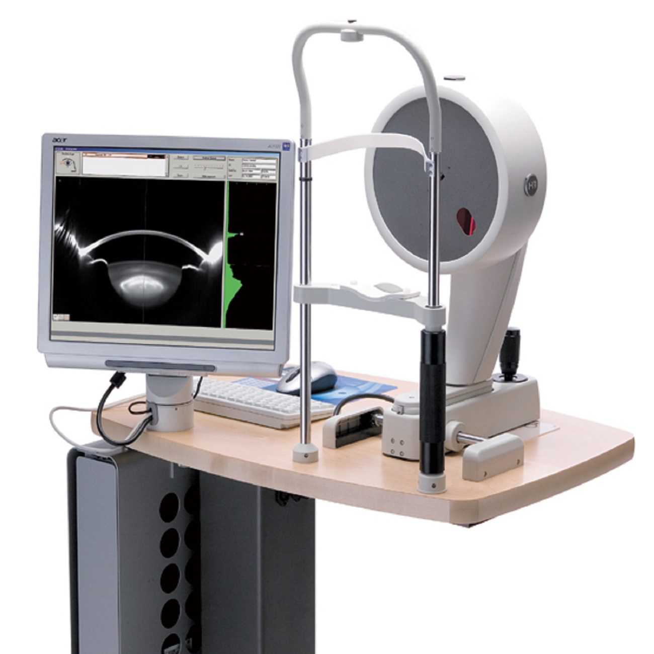
(17) PENTACAM HR
High resolution cross sectional images of the anterior segment
Axial and tangential topography maps
Total corneal refractive power
Pachymetry maps
Anterior and posterior corneal elevation maps
Zernike corneal wavefront analysis
Anterior chamber angle measurement
Anterior chamber depth and volume measurements
3D Lens Densitometry and Pentacam Nucleus Staging (automatic classification of lens opacity)
Corneal Optical Densitometry for assessing and quantifying corneal opacification
Fast Screening Report to quickly identifying abnormalities found in common pathologies
Cataract Pre-op Display for premium IOLs
Holladay Report with equivilent K readings
Keratoconus detection and staging
Belin/Ambrosio Enhanced Ectasia
Contact Lens Fitting with simulated fluorescein image
Various comparison displays including Compare 2 Scheimpflug
Network ready - Up to 50 workstations included
